By Jo Adaway
Consultant Clinical Scientist, Biochemistry
April 2023
It is estimated that 26-30% of adults in the UK have hypertension, equating to around 13.2 million people in the UK. Up to 10% of the hypertensive population have a secondary cause of their hypertension, affecting around 1.3 million patients. Identification of affected patients will allow optimal clinical response to be achieved with the specific therapy.
The following groups of patients should be screened for secondary causes of hypertension:
- Young onset hypertension (before the age of 40) NG136
- Drug resistant hypertension
- Sudden deterioration in blood pressure control
- Hypertensive emergency
- Clinical clues
- Muscle weakness/tetany, cramps, or arrhythmias (Primary hyperaldosteronism)
- Rapid onset acute pulmonary oedema (renal artery stenosis)
- Sweating, palpitations, or frequent headaches (phaeochromocytoma/paraganglioma)
- Snoring or daytime sleepiness (obstructive sleep apnoea)
Renal disorders such as CKD, diabetic nephropathy or polycystic kidney disease are the most common cause of secondary hypertension. Other causes include vascular disorders such as renal artery stenosis, drugs and medications, obstructive sleep apnoea, pregnancy and endocrine disorders. Clinical examinations and routine biochemical tests can indicate which are the most likely secondary causes and signpost which further investigations could be helpful.
Renal disease
Initial investigations for CKD should include eGFR and urine albumin:creatinine ratio (NG203). Early morning urine samples are recommended for the albumin:creatinine ratio. These tests are also recommended to be carried out annually in type 1 diabetics to identify diabetic nephropathy. If polycystic kidney disease is suspected, renal ultrasound is the diagnostic procedure of choice.
Primary hyperaldosteronism
Primary hyperaldosteronism or Conn’s syndrome is the cause of up to 23% of cases of drug resistant hypertension (Endocrine Society Clinical Practice Guidelines). Aldosterone:renin ratio (ARR) is the first line test for the diagnosis of primary hyperaldosteronism. An EDTA plasma sample is needed for this test; samples are stable for up to 24 hours at room temperature so can be taken using routine phlebotomy services and sent in the usual transport to the laboratory for processing. The samples are analysed using mass spectrometry, which is a highly specific method, and results should be available within two weeks.
Patients should be encouraged not to restrict sodium intake prior to the test, and if the patient is hypokalaemic, this should ideally be corrected before the sample is taken. The Endocrine Society recommend discontinuation of aldosterone antagonist (spironolactone and epleronone) and diuretics for 4 weeks prior to the test if possible; other drugs should not cause diagnostic confusion in the majority of cases. However, if it is not clinically possible to withdraw these drugs, the samples should still be taken and the use of these drugs indicated in the clinical details, to enable proper interpretation of the results.
If the ARR is 1000, the primary hyperaldosteronism is unlikely. ARR >2000 with aldosterone >400 pmol/L is indicative of primary hyperaldosteronism, and any patients with these results should be referred to endocrinology for further investigations. Repeat samples will be requested if the ARR is 1000-2000 or is >2000 but the aldosterone <400 pmol/L. If these results are confirmed on repeat, a referral to endocrinology is again recommended.
Phaeochromocytoma/paraganglioma
This is a rarer cause of hypertension, but is thought to affect around 0.6% of hypertensive patients, and the diagnosis is more likely if the patient also has symptoms such as sweating, palpitations or frequent sweating. Plasma metanephrines (metanephrine, normetanephrine and 3-methoxytyramine) are metabolites of catecholamines and are produced by the action of the enzyme catechol-O-methyl transferase on adrenaline, noradrenaline and dopamine. This enzyme is present within tumour cells, which is the reason why metanephrines show superior sensitivity to catecholamines in the diagnosis of PPGL (Eisenhofer et al, 1998). Plasma metanephrines are more sensitive than urine metanephrines for PPGL diagnosis (97% for plasma vs 90% for urine) (Sawka et al 2003) which is why plasma metanephrines is the test offered for the investigation of PPGL in this Trust.
Plasma metanephrines are unstable and must be received in the laboratory within 1 hour of collection, where they are centrifuged, the plasma is separated from the blood cells, and frozen prior to analysis. For this reason, samples must be collected in the hospital and the test is not available for GPs to request. It is recommended that patients with suspected PPGL are referred to endocrinology for investigation. If there is a specific reason why this is not possible, contact the Duty Biochemist (291 2136 for Wythenshawe and Trafford, 701 8891 for MRI and North Manchester) who will be able to advise.
Cushing’s syndrome
Cushing’s syndrome is a very rare cause of secondary hypertension (<0.1%) of cases. Symptoms of Cushing’s include central weight gain, easy bruising, buffalo hump, round puffy face, proximal muscle weakness and abdominal striae. If Cushing’s syndrome is clinically suspected, there are several tests that can be performed to identify if the patient has hypercortisolism; however random serum cortisol is not useful for this.
- Urine free cortisol – at least 2 24-hour urine collections in plain bottles. The collections must be accurate – if not all the urine passed in the 24 hour period is collected, the result reported would be falsely low, and if the collection is more than 24 hours’ worth of urine, the result would be falsely high.
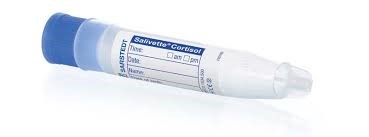
- Late night salivary cortisol – this is often tolerated well by the patients. Contact the lab to arrange for saliva collection devices (salivettes) to be sent to your surgery. At midnight, the patient should gently chew the swab for 1-2 minutes then replace it in the collection device. The sample can then be returned to the surgery or directly to the lab. Samples should not be taken within 30 minutes of eating, drinking or tooth brushing.
- Overnight dexamethasone suppression test – the patient takes 1 mg of dexamethasone at 11 PM and a blood sample for cortisol is taken at 9 AM the next morning. In normal subjects, the cortisol will suppress to <50 nmol/L. If the cortisol is above 50 nmol/L, the lab will add dexamethasone analysis onto the sample to confirm adequate absorption and metabolism of dexamethasone. The dexamethasone and samples must be taken at the correct times to ensure accurate interpretation of results is possible.
If any of these tests is abnormal, referral to endocrinology is recommended for further investigations.
Thyroid disease
Hypo- and hyperthyroidism can both rarely cause secondary hypertension, although the mechanism is poorly understood. Common symptoms that may indicate hypothyroidism include tiredness, cold intolerance, weight gain, constipation and muscle weakness; symptoms of hyperthyroidism are weight loss, mood swings, difficulty sleeping, heat sensitivity, palpitations or neck swelling. Thyroid function tests (TSH and fT4) should be requested to investigate thyroid disorders. In primary hyperthyroidism, the TSH will be low with a raised fT4 or fT3 (the Duty Biochemist will add fT3 onto samples as necessary depending on the pattern of results). In primary hypothyroidism, the TSH will be raised and the fT4 may be low or at the lower end of the reference range. It is important to include accurate clinical details with all thyroid requests to ensure the proper interpretation can be added to the results.
There are many causes of secondary hypertension. If you suspect your patient may have a secondary cause of their high blood pressure and you are unsure as to the appropriate investigations, contact the Duty Biochemist for advice:
- MRI/North Manchester: 701 8891 or Dutybiochemist.orc@nhs.net
- Wythenshawe/Trafford: 291 2136 or mft.biochemistry.wythenshawe@nhs.net
References
NICE Guideline 136. Hypertension in adults: diagnosis and management.
NICE Guideline 203. Chronic kidney disease: assessment and management.
Funder J et al. The Management of Primary Hyperaldosteronism: Case Detection, Diagnosis and Treatment: An Endocrine Society Clinical Practice Guideline. JCEM 2016; 101: 1889-1916.
Eisenhofer G et al. Plasma metanephrines are markers of pheochromocytoma produced by catechol-O-methyl transferase within tumors. JCEM 1998; 83:2175-2185.
Sawka AM et al. A comparison of biochemical tests for pheochromocytomas: measurement of fractionated plasma metanephrines compared with the combination of 24-hour urinary metanephrines and catecholamines. JCEM 2003; 88:553-8.
 In this section
